
Decoding Neurogenetic Answers- Case #3

Patient Clinical Information: The proband is a 6 year-old female with a primary clinical phenotype of intellectual disability. Additionally, it was noted that the proband has proximal and distal weakness, muscle atrophy, and severe hypotonia. She has absent osteotendinous reflexes and severe hypoacusia (hearing loss). A thin corpus callosum was also noted on the patient’s brain MRI.
Family History: None reported.
Testing Ordered: MNG Exome™ Trio Sequencing + mtDNA
Genetic Findings: Through exome sequencing, the proband was found to have an apparent homozygous single nucleotide C duplication in the beta IV spectrin gene, SPTBN4. The proband’s father was shown to be heterozygous for the same C duplication, and the mother appears to be wild type, making this an interesting discovery. When copy number data was analyzed, it was discovered that both the proband and the mother have a heterozygous deletion in the SPTBN4 gene that spans exons 6-11. The duplication variant previously noted is in exon 10. These findings explain why the proband appears to be homozygous for the duplication variant.
Outcome: Based on the sequencing and copy number findings, this lead to a diagnosis of Congenital Myopathy with neuropathy and deafness, or CMND. This syndrome is a recently described autosomal recessive condition that is caused by mutations in the SPTBN4 gene. The beta IV spectrin protein is expressed in brain, peripheral nervous system, pancreas and skeletal muscle. CMND is characterized by severe hypotonia, muscle atrophy, areflexia and hearing loss. To date in the literature, only seven individuals that harbor either homozygous or compound heterozygous putative loss of function variants in the SPTBN4 gene have been reported. By combining whole exome sequencing and copy number analysis, MNG was able to help identify the genetic cause of the patient’s symptoms that otherwise would have gone undetected.
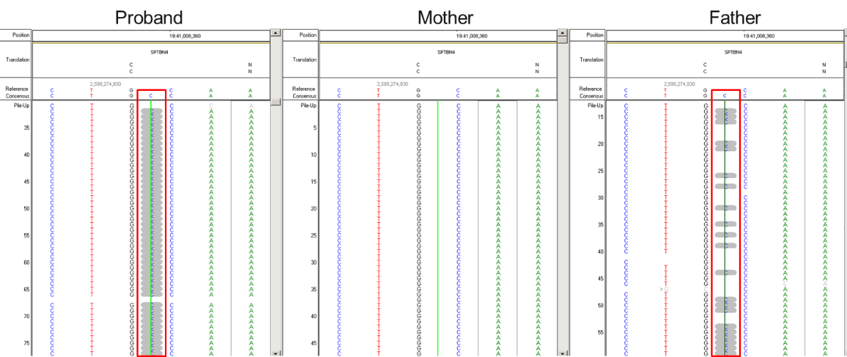
Figure 1: This image shows comparative sequencing reads from the proband and both parents. The red box highlights the C duplication found in SPTBN4 in both proband, apparently homozygous, and father, heterozygous. The green reference line seen in the mother’s sequencing reads shows no reads with a C duplication.
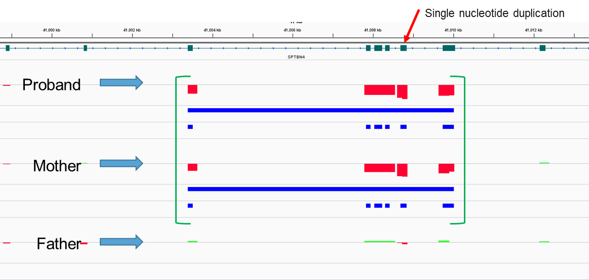
Figure 2: Copy number analysis visualization of proband, mother, and father. The green brackets show the area of interest between exons 6-11, and a heterozygous deletion of those exons in both the proband and mother. The arrow shows exon 10, where the previously noted duplication was detected through sequencing analysis.
Reported by: Heather Marton, PhD, Reporting, Laboratory Liaison